Eye health is a crucial component of overall well-being, yet even a seemingly minor infection can compromise both comfort and vision. Eye infections are common across all age groups and may present with symptoms ranging from mild irritation to severe inflammation that threatens sight. Recognizing the early signs, understanding the underlying causes, and seeking timely medical evaluation are essential for preserving long-term ocular integrity.
Defining an Eye Infection
An eye infection occurs when pathogenic microorganisms, such as bacteria, viruses, fungi, or parasites, invade ocular tissues or adjacent structures. The infection may involve the eyelids, conjunctiva, cornea, or intraocular spaces. Depending on its origin and severity, an infection can cause discomfort, disrupt normal tear film function, or, in advanced cases, lead to permanent visual impairment. While the term “eye infection” is often associated with conjunctivitis (pink eye), the category encompasses a broad spectrum of conditions, each requiring distinct diagnostic and therapeutic approaches.
What Causes Eye Infections? Epidemiology and Risk Factors
Eye infections represent a significant portion of ocular complaints seen in both primary care and optometry clinics.
In the United States, over a million visits annually are attributed to infectious eye diseases. Contact lens wearers are at particularly elevated risk, especially when lenses are worn overnight, inadequately disinfected, or exposed to contaminated water. Other risk factors include poor eyelid hygiene, compromised immune function, and previous ocular trauma or surgery. Common eye infections are:
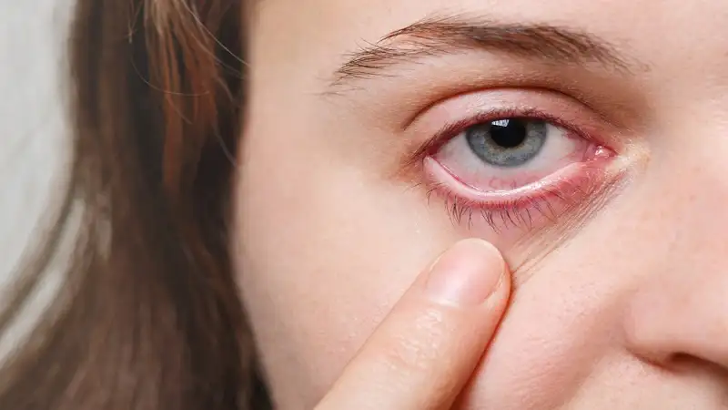
Conjunctivitis (Pink Eye)
Conjunctivitis involves inflammation of the conjunctiva, the transparent membrane lining the eyelid and covering the sclera. It manifests as redness, itching, tearing, and discharge that may cause the eyelids to stick together upon waking.
Conjunctivitis can be:
- Viral: Typically secondary to upper respiratory infections and spread via direct contact.
- Bacterial: Commonly caused by Staphylococcus aureus, Streptococcus pneumoniae, or Haemophilus influenzae, often producing thick mucopurulent discharge.
- Allergic: Triggered by airborne allergens such as pollen or dust mites, leading to bilateral itching and watery discharge.
For management strategies, refer to our comprehensive resource on how to get rid of pink eye overnight.
Keratitis
Keratitis denotes inflammation or infection of the cornea and can rapidly progress if untreated. Microbial keratitis is most frequently associated with contact lens misuse, while non-infectious keratitis may result from trauma, UV exposure, or systemic inflammatory disorders. Clinical features include ocular pain, photophobia, blurred vision, and excessive tearing. Corneal ulceration and scarring may occur in advanced cases, necessitating urgent ophthalmologic care.
Blepharitis
Blepharitis is characterized by chronic inflammation of the eyelid margins, often involving the meibomian glands.
It may be bacterial, seborrheic, or demodex-related. Patients commonly report itching, burning, crusting at the lash line, and a sensation of dryness or grittiness. Long-term management includes meticulous lid hygiene, warm compresses, and, in persistent cases, topical or systemic antibiotics.
Stye (Hordeolum)
A stye represents an acute infection of the eyelid’s oil gland or hair follicle, typically caused by Staphylococcus aureus. It presents as a localized, tender, erythematous nodule along the lid margin. Although self-limiting, warm compresses and proper eyelid hygiene expedite resolution.
Persistent or recurrent styles may warrant evaluation for underlying blepharitis or chronic bacterial colonization.
Cellulitis
- Periocular cellulitis is an infection of the eyelid and surrounding soft tissue, often secondary to trauma or sinus infection.
- Preseptal cellulitis affects the superficial tissues anterior to the orbital septum and generally responds to oral antibiotics.
- Orbital cellulitis, by contrast, extends posteriorly into the orbital cavity, posing a serious threat to vision and requiring prompt hospitalization and intravenous therapy.
Uveitis
Uveitis involves inflammation of the uveal tract, including the iris, ciliary body, and choroid. It may arise from autoimmune disorders, systemic infections, or ocular trauma. Symptoms include ocular pain, photophobia, blurred vision, and redness around the iris (ciliary flush). Early intervention is essential to prevent complications such as glaucoma or macular edema.
Endophthalmitis
Endophthalmitis is a rare but devastating intraocular infection, most commonly occurring after surgery, trauma, or intravitreal injection. Rapid onset of severe pain, visual loss, and marked redness necessitates immediate ophthalmic intervention. Delay in treatment can result in irreversible blindness.
Etiology and Contributing Factors
Ocular infections are typically initiated by microbial entry through breaks in the ocular surface or via contaminated contact lenses, cosmetics, or hands. Common pathogens include:
- Bacteria: Staphylococcus aureus, Streptococcus pneumoniae, Pseudomonas aeruginosa
- Viruses: Herpes simplex virus, adenovirus
- Fungi: Candida and Fusarium species
- Parasites: Acanthamoeba, often associated with exposure to non-sterile water while wearing contact lenses
Environmental exposure, reduced tear film quality, and underlying systemic illness can all compromise the eye’s natural defense mechanisms, increasing susceptibility to infection.
Recognizing the Clinical Presentation
The presentation of an eye infection can vary depending on the affected tissue and pathogen. Key clinical indicators may include:
- Conjunctival redness or hyperemia
- Foreign body sensation or irritation
- Excessive tearing or discharge (clear, mucous, or purulent)
- Eyelid swelling and tenderness
- Photophobia (light sensitivity)
- Blurred or fluctuating vision
- Crusting along the eyelashes upon waking
Any sudden onset of pain, swelling, or visual disturbance should prompt immediate examination by an eye care professional.
Diagnostic Approach and Treatment Modalities
Clinical Evaluation
Diagnosis is established through comprehensive ocular examination, including slit-lamp assessment, fluorescein staining, and, in certain cases, microbial cultures or PCR testing to identify the causative organism.
A detailed history of contact lens use, recent illness, or systemic disease assists in determining the likely etiology.
Therapeutic Management
Treatment protocols are determined by the type and severity of infection:
- Bacterial infections are managed with topical or systemic antibiotics, depending on tissue involvement.
- Viral infections often resolve spontaneously; however, antiviral therapy may be indicated for herpes-related keratitis or uveitis.
- Fungal infections require prompt antifungal therapy and close monitoring.
- Allergic or inflammatory causes respond well to antihistamines, mast cell stabilizers, or corticosteroid drops under medical supervision.
During recovery, patients should avoid contact lenses and practice stringent hand and eyelid hygiene to prevent reinfection.
When to Seek Immediate Medical Attention
While mild irritation may not constitute an emergency, certain symptoms warrant urgent evaluation:
- Severe ocular pain or swelling
- Rapid decline in visual acuity
- Pronounced light sensitivity
- Persistent redness unresponsive to home care
- Signs of systemic infection such as fever or malaise
Early medical intervention minimizes the risk of complications, including corneal scarring and permanent vision loss.
Preventive Strategies
Maintain Rigorous Hygiene
Hand hygiene remains the most effective preventive measure. Avoid touching or rubbing the eyes unnecessarily, and refrain from sharing towels, cosmetics, or personal care items.
Contact Lens Compliance
Adhere strictly to lens cleaning, storage, and replacement guidelines. Avoid overnight wear unless explicitly approved by your eye care provider, and never expose lenses to water.
Protective Measures
Use appropriate eye protection during activities involving potential exposure to dust, chemicals, or debris.
Routine Eye Examinations
Regular comprehensive eye exams play a vital role in identifying early signs of infection or inflammation, allowing for timely intervention before vision is compromised.
The Clinical Perspective
Eye infections are not only a source of discomfort but can also have lasting implications for ocular health if neglected. Recognizing early warning signs, maintaining strict hygiene practices, and pursuing prompt, professional evaluation are fundamental to preserving vision and ocular function. Through consistent care and informed awareness, the risk of infection can be significantly reduced, ensuring the eyes remain clear, comfortable, and healthy for years to come.